The case documentation below describes the surgical treatment of tarsometatarsal (TMT) osteoarthritis II and III. The 77-year-old patient has dorsalis pedis stress and rolling pain in the tarsal region. The quality of life is increasingly limited, the walking distance shortened due to pain. After multiple successful infiltrations that provided short-term pain relief, the patient opts for arthrodesis of TMT joints II and III, each with a human bone screw graft. After surgery, the patient was immobilized in a hallux shoe for six weeks.
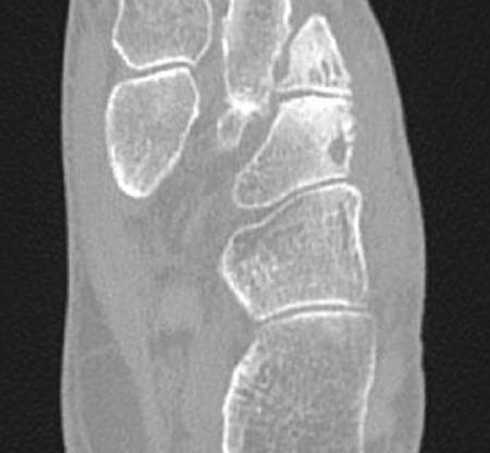
CT preoperatively
For verification of osteoarthritis
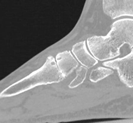
CT preoperatively
Shows significant osteoarthritis in the TMT joint
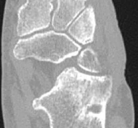
CT preoperatively
Shows significant osteoarthritis in the TMT joint
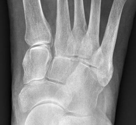
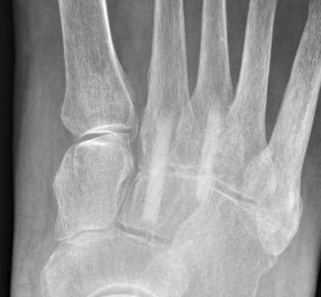
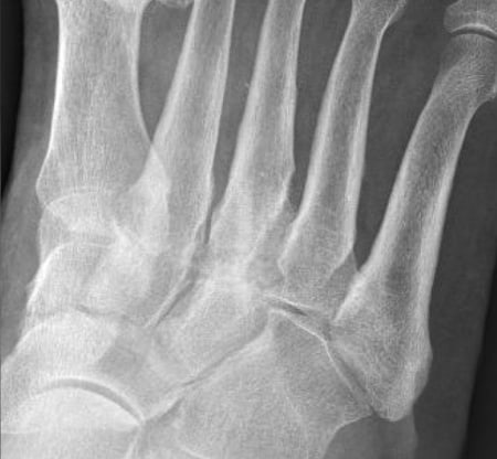
X-ray image preoperatively
The control X-ray clearly shows the arthrosis
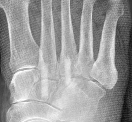
X-ray postoperatively
Treatment of osteoarthritis with Shark Screw® grafts 5.0mm
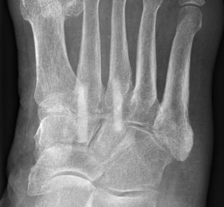
6 weeks postoperatively
Optimal position of the grafts, beginning of the arthrodesis
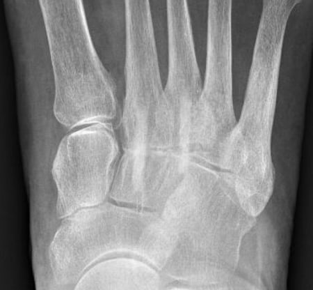
3 months postoperatively
Good build-up of the arthrodesis, stable position of the Shark Screw® grafts, no osteolysis
6 months postoperatively
Good build-through, the grafts are more and more remodeled into their own bone
1 year postoperatively
Complete non-reactive arthrodesis of TMT joints II and III, the Shark Screw® grafts barely visible, patient-derived bone has emerged
OP technique & course
After imaging the TMT II and III joints under image intensifier control, the joint space is held open with spreading forceps and the remaining articular cartilage is removed with a sharp spoon. The underlying sclerotic bone is tapped with a thin drill. After the two joints can be adjusted well to each other, the KD is placed under image converter control. Additional insertion of a temporary KD for stable fixation of the joints for the time of drilling and tapping. Knochen in diesem Fall extrem weich. The thick graft of 5mm facilitates arthrodesis because the fine thread has a very good hold in the thin bone, which is drilled into almost tangentially. Very stable osteosynthesis. The joint space completely closed. A metallic lag screw would tear out in the soft bone. The pressure exerted on the joint by the surgeon is perfectly sufficient to optimally align the joint surfaces.
Advantages with this surgery
no metal, head is placed introperatively at bone level and thus the surrounding soft tissues are spared. Therefore, less pain for the patient. Da der Knochen sehr weich war und große Zysten aufwies, bestand nach der Transplantation der 5,0mm dicken Shark Screw® eine sehr verlässliche und stabile Knochenbrücke. Metal screws and metal plates wear very heavily in this area, arroding the tendons. The hold of metallic screws would have been questionable.
Subjective patient satisfaction
Patient had no pain in the postoperative course, full weight-bearing capacity after 6 weeks. Ready-made shoes fit again after 6 weeks. She was so pleased with the initial surgery that she had the second side operated on 3 months later.