The following case documentation describes the medical course of cartilage-bone necrosis at the medial femoral condyle of the left knee joint. Since the 41-year-old patient suffers from a very large (2×3.5cm) and deep (8-10mm) cartilage-bone necrosis, a “fresh-frozen” cartilage-bone graft is performed. The necrotic areas are freshened and drilled into. An impression of the defect is then taken with bone wax, the graft is remade to the size of the impression using a hallux saw, and fixed with four human bone screw grafts.
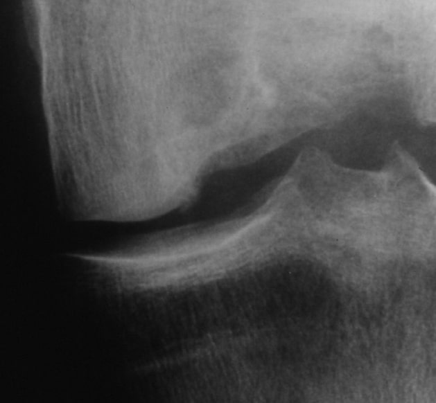
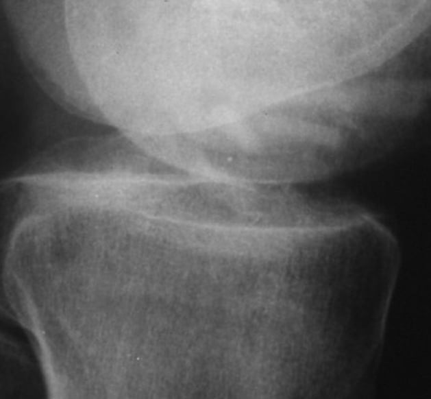
X-ray image preoperatively
Deep defect at the medial femoral condyle of the left knee joint
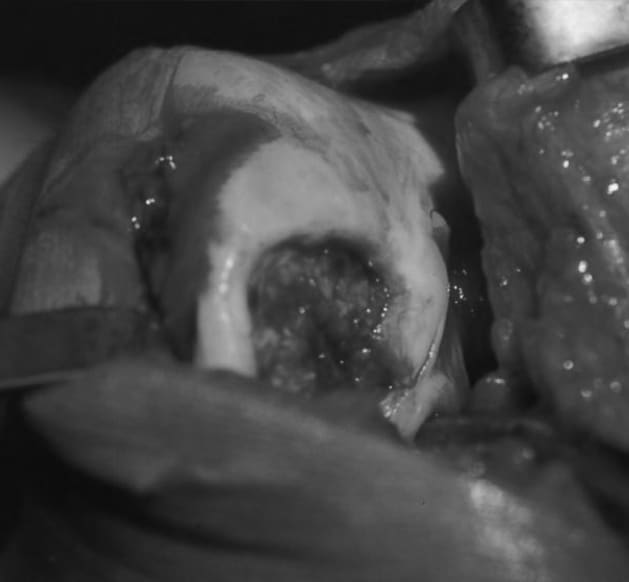
Situs intraoperativ
Before removing the necrotic tissue and before milling and drilling
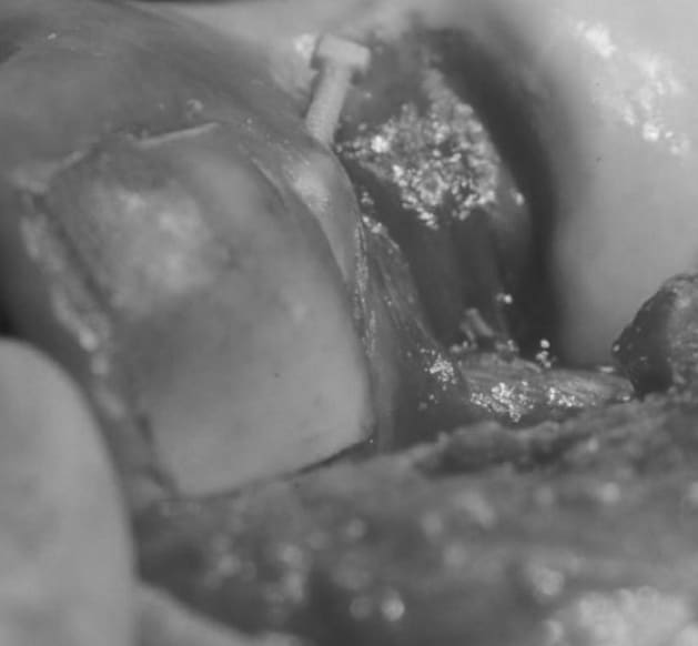
Situs intraoperatively
"Fresh-Frozen" graft is inserted. Bone screw graft in the intercondylar notch
Situs intraoperatively
6 months postoperatively Bone screw grafts are well visible. No osteolysis visible
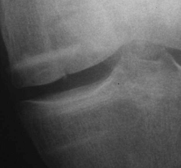
6 months postoperatively
X-ray control lateral. Graft has grown in.
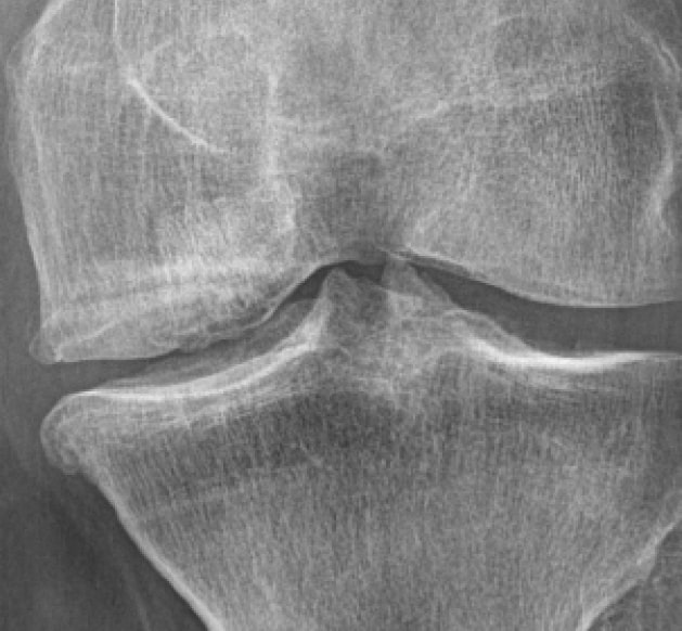
16 years postoperatively
Moderate medial osteoarthritis with good cartilage-bearing layer, patient is symptom-free
Clinical case of Dr. Klaus Pastl, Linz in Austria. “Fresh-frozen” cartilage-bone grafting at the medial femoral condyle with four bone screw grafts. (documentation period 1995-2011) © surgebright GmbH