The following case report describes the medical course of osteochondritis dissecans (OD). The 26-year-old patient has been complaining of load-dependent pain in the knee joint for months. A few days before the examination, the patient also suffers from symptoms of entrapment and restricted movement. The radiograph and MRI show OD with a free joint body and empty mouse bed. During surgery, the free joint body – despite being thin and subchondral – is grasped and refixed into its bed with the help of the bone screw graft. The protruding part of the graft is trimmed back to the subchondral bone level using a bone reamer.
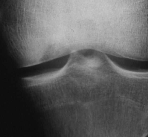
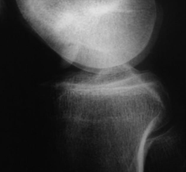
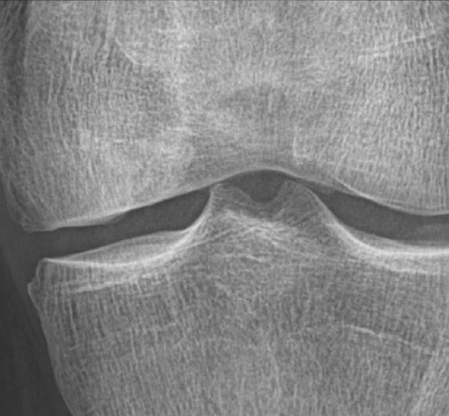
X-ray image preoperatively
Deep osteochondritic defect. Free joint body lies in the mouse bed
X-ray postoperatively in plaster
Sagittal Z-shaped shortening of the ulna and osteosynthesis with five bone screw grafts.
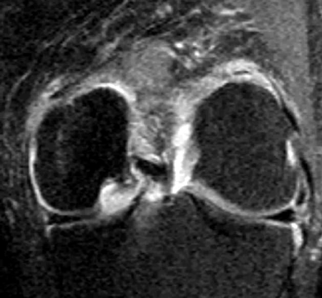
MRI preoperative
Free joint body lies intercondylar. Clear joint effusion and deep and free mouse bed visible.
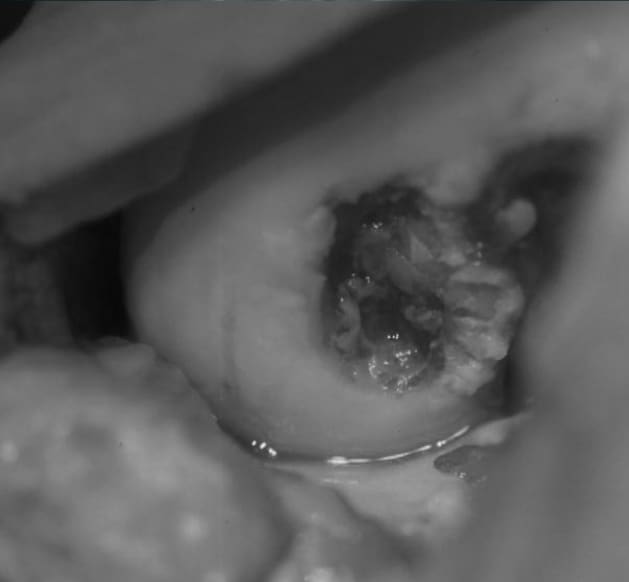
Situs intraoperatively
Punched out defect. Mouse bed is freed from necrotic material, tapped and relined with autologous bone
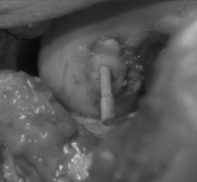
Situs intraoperatively
Free joint body is refixed with bone screws graft. Protruding head is shortened
11 years postoperatively
The patient is symptom-free
Clinical case of Dr. Klaus Pastl, Linz in Austria. Refixation of a free joint body with a human bone screw graft. (documentation period 1996-2007) © surgebright GmbH